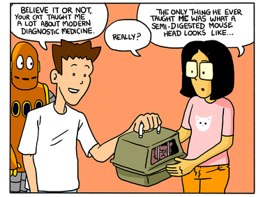
Tim is taking care of Pickle, Rita's cat while she is gone and Moby tries to scan his body. Tim doesn't allow Moby to scan Pickle because he thinks he is trying to harm Pickle instead of scanning Pickle. Tim reads a letter and teaches you about body scans. At the end, Tim figures out that Moby was just trying to just scan Pickle's body and finds a Thundercat action figure that Tim owned. Then, they take Pickle to the vet.
(Moby is chasing his kitty Pickles until Tim stops him.)
Tim: Moby! (Pickle jumps on him.) You know, I promise Rita I'd take care of Pickle while she's away, so you can't run around shooting lasers at him. (Moby scans Pickles with his own laser eyes, and Pickle runs away.) And you can't…do whatever that was either.
FYI's[]
Trivia[]
In May of 2006, a researcher in San Antonio, Texas developed a miniature version of the CT scan that can be used to see inside living human embryos! Dr. Charles Keller of the University of Texas Health Science Center developed the technique, which will be able to spot the earliest signs of abnormalities, cancers, and genetic defects.
In January 2005, a team of scientists, working alongside the Egyptian government, removed the 3,500-year-old mummy of Tutankhamen (King Tut) from its tomb in Egypt’s Valley of Kings. They took it to a mobile CT scanner that had been brought near the site, and performed a 15-minute CT scan on the body (pictured). After examining the images, the scientists determined that contrary to rumor, King Tut definitely did not die of a blow to the head. He did, however, have a badly fractured leg, although scientists disagree over whether King Tut suffered the injury when he was alive, or if the break occurred after he died.
Godfrey Hounsfield and Allan Cormack, inventors of the CT scan, won the Nobel Prize in Medicine 1979. Paul Lauterbur and Sir Peter Mansfield were awarded the Nobel Prize in Medicine in 2003 for their work with MRI machines.
In Depth[]
How, exactly, does MRI work? Well, an MRI machine contains incredibly powerful magnets. In fact, before having an MRI, you have to remove pens, keys, and loose change from your pockets. This is because the magnetic field is so strong that it can turn these objects into dangerously fast moving objects!
Hydrogen atoms exhibit interesting behavior when they’re placed in a magnetic field—their protons tend to line up in the direction of the field. So, before it does anything else, the MRI machine causes the hydrogen atoms in your body to line up. Then, it sends out a pulse of radio waves, pinpointed to the area of the body that needs to be examined.
These waves have a frequency that’s specific to hydrogen, and, like all electromagnetic waves, they contain energy. The hydrogen protons absorb this energy, which causes them to spin in a different direction. When the pulse is turned off, the hydrogen atoms release the energy they absorbed. The energy they release is picked up and sent to a computer, which uses that information to form a picture.
In Practice[]
Nuclear medicine, the use of radioactive chemicals to help in medical imaging, has become a very important tool for doctors. Radioactive chemicals are injected into the patient and then detected by machines like a PET scanner. The chemicals contain nuclei from radioactive elements.
Here is a list of the most commonly used elements in nuclear medicine! Note that the numbers refer to a particular isotopes’ mass number—the combined number of protons and neutrons in the atomic nucleus.
Technetium-99
Iodine-123 and -131
Thallium-201
Gallium-67
Fluorine-18
Indium-111
Xenon-133
Krypton-81
Arts and Entertainment[]
What does the Beatles’ Sergeant Pepper’s Lonely Hearts Club Band have to do with body scan technology? More than you might imagine.
One of the two inventors of the CT scan was a British researcher named Godfrey Hounsfield, who worked for a company called Electronic and Musical Instruments, or EMI, for short. EMI was involved in a lot of high-tech stuff, and Hounsfield worked on their radar and guided weapons systems before helping them build the first all-transistor computer in Great Britain. He began work on what would become the CT scanner in 1967; after five years of work, it debuted in 1972. The first CT scanners were all built by EMI; in fact, the very first one was called the “EMI scanner.”
At that time, the Beatles were the most popular, best-selling musical group in the world. They had grossed an estimated $98 million worldwide by May 1967, and the album they released in June of that year, Sgt. Pepper, stayed at No. 1 for 27 weeks in Britain and 15 weeks in America.
What’s the connection? Well, EMI just happened to be the Beatles’ record company. EMI needed a lot of money to fund Hounsfield’s cutting-edge research into computed tomography, and it had this money on hand due to the Beatles’ unmatched success. Some people have even claimed that the CT scan is the Beatles’ “greatest legacy,” since it never would have been built without the money they made. Goo goo ga joob!
Real Life[]
If you go to the hospital with a painful broken arm, the first thing a doctor usually does is order x-rays. These scans show where the problem is and how severe it is, so that the doctor can fix it. But what if the source of your pain is, say, an overwhelming feeling of hopelessness? What kinds of tests can a doctor order for that?
Feeling hopeless is just one of many ways that depression can affect a person. One patient may report a loss of appetite and mood swings, while the next may describe excessive sleepiness and feelings of guilt. This variability has made depression tricky for doctors to diagnose and choose the best treatment.
Right now, doctors largely rely on patients’ own descriptions of their symptoms, or signs of illness. Instead of ordering a scan or test, doctors have their patients complete questionnaires about how they feel. While this information is helpful, it is not enough. Doctors often have to go through a process of trial-and-error when treating patients for depression.
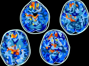
To help doctors help their patients with depression, scientists are studying the brain itself. Some of the most exciting discoveries have come from scans that reveal what is happening inside the brain, kind of like how x-rays reveal our bones. Functional magnetic resonance imaging (fMRI) scans show which areas of the brain are active under various conditions. For example, depending on whether you have just watched a video of a cute bunny or one of an angry bear, different parts of the brain may light up with activity. You can probably guess which video would spark more activity in the part of the brain that processes fear.
The brains of depressed people and non-depressed people look different in fMRI scans, according to a major study published in 2017. Even more importantly, scientists found patterns in the fMRI scans of depressed patients who reported particular symptoms. One key discovery was that patients who reported feeling abnormally tired showed different patterns of brain activity than patients who reported having difficulty feeling pleasure. And patients in one group were more likely to benefit from a particular treatment than the other.
Findings like these suggest that one day, doctors will not have to keep resorting to trial and error when treating depression. With the help of science, treating depression will hopefully become as straightforward as treating a broken arm.


